An MCQ based case discussion by
Dr. Smitirupa Mishra , Consultant Pathologist, Sparsh Hospital , Bhubaneswar, Odisha
CASE HISTORY:
A 50-year-old male presents with pain abdomen and dyspepsia. Intraoperatively, appendicular perforation with psoas abscess is noted. Appendectomy specimen received for HPE
See below the Histopathology images based on which MCQs are framed.
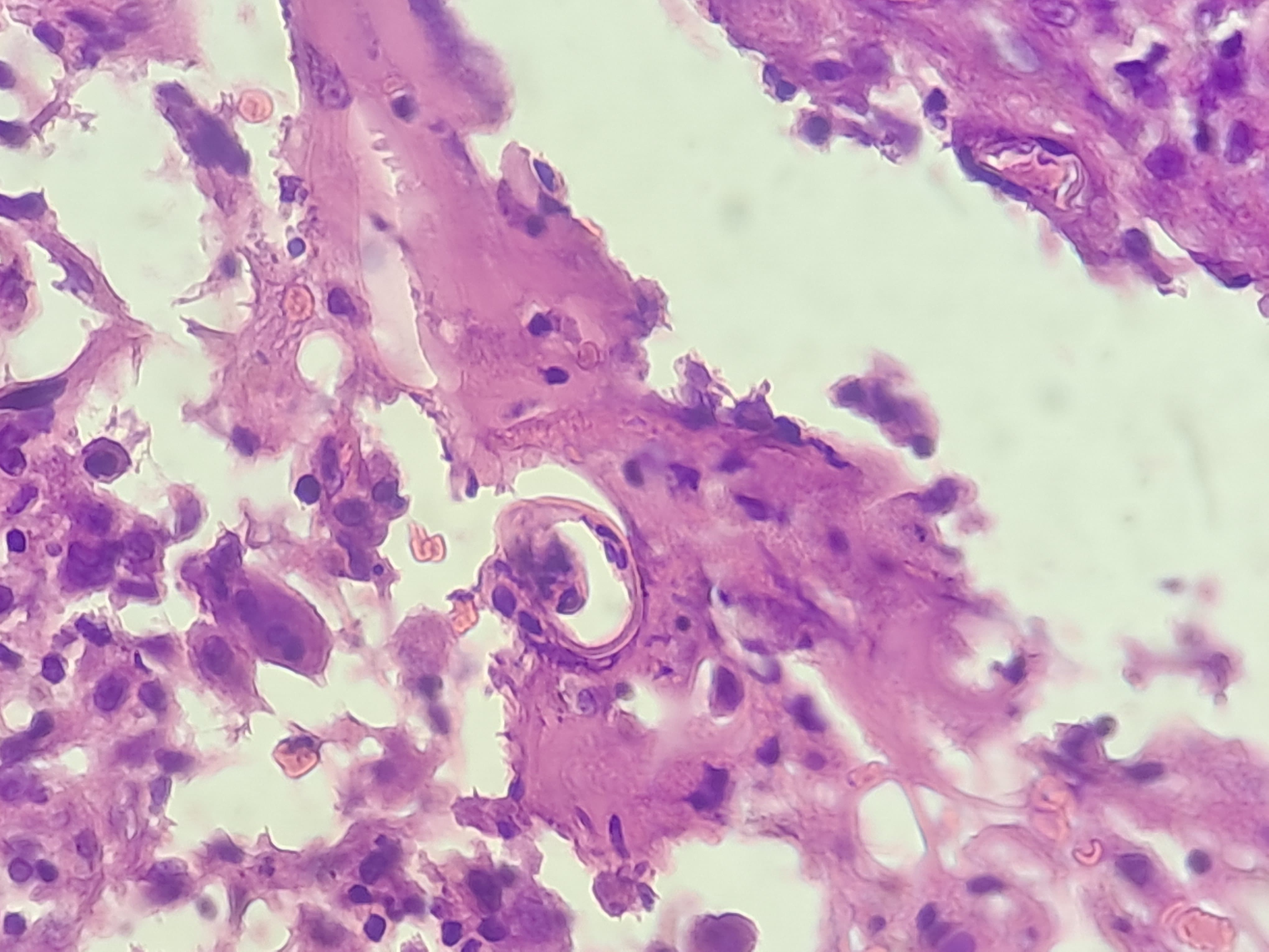
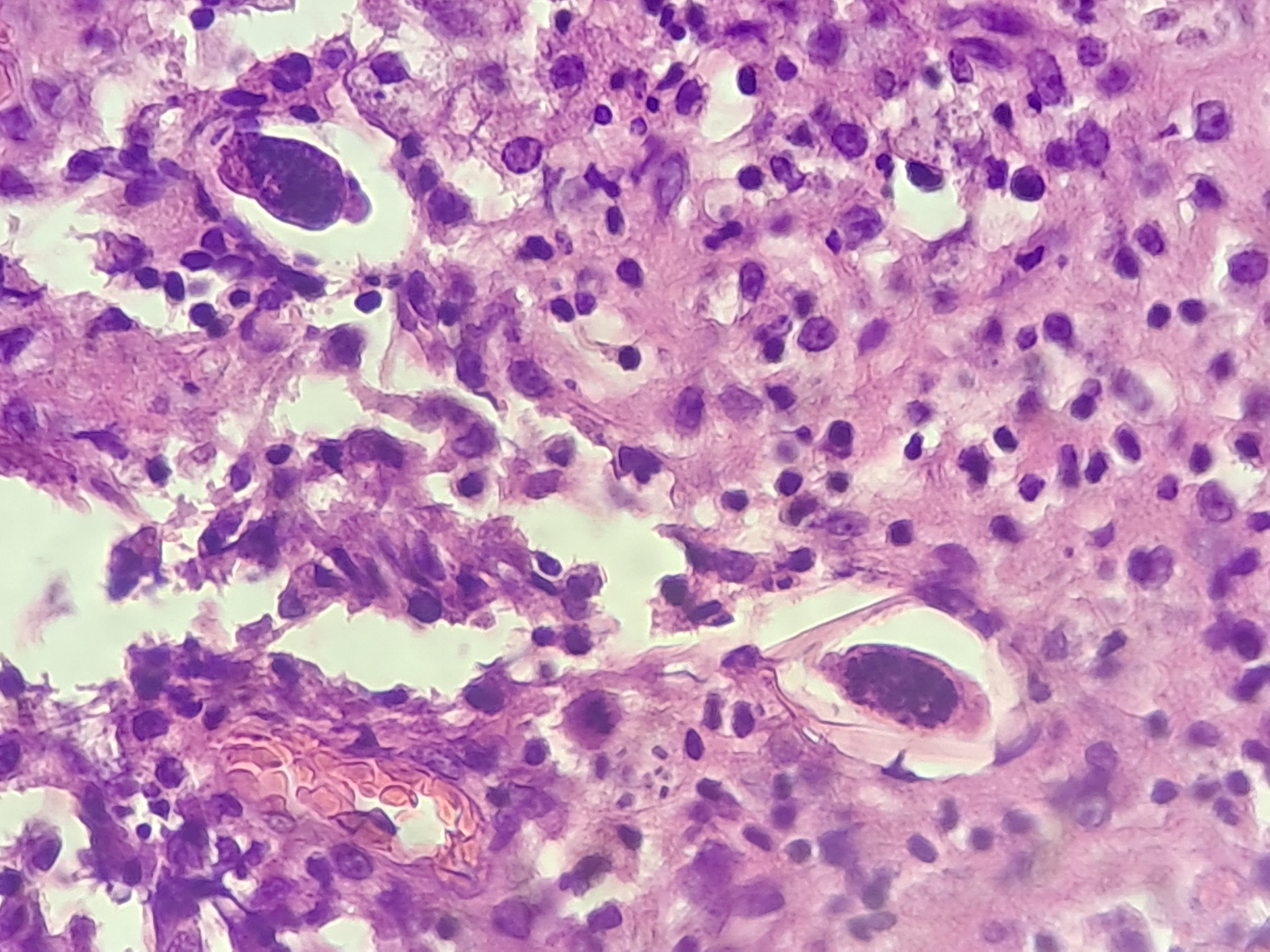
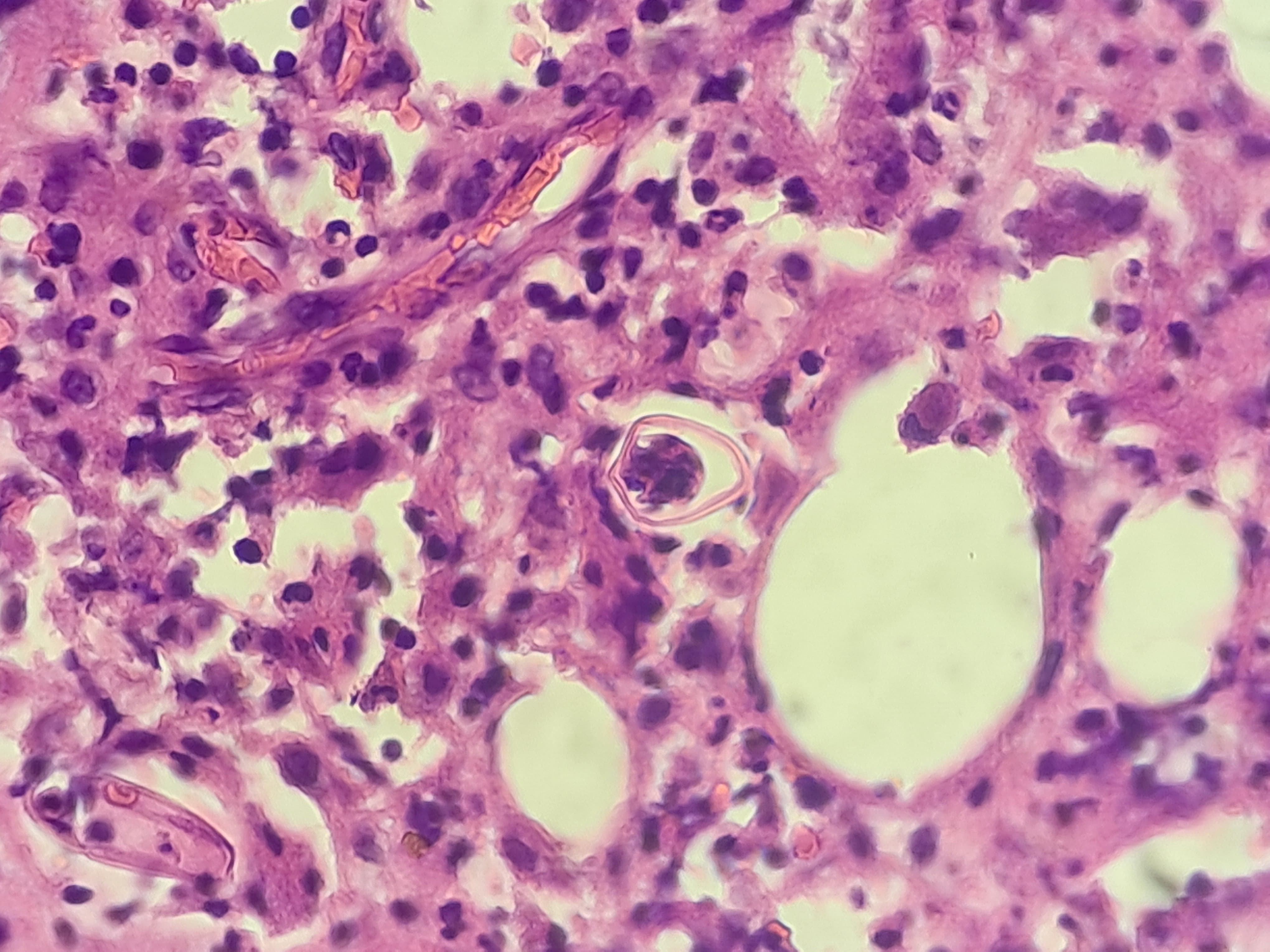
Q1. Identify the organism seen in the appendiceal lumen in the given histopathology image:
A. Ascaris lumbricoides
B. Enterobius vermicularis
C. Taenia solium
D. Trichuris trichiura
Q2. The most characteristic microscopic feature of this organism is:
A. Barrel-shaped eggs with bipolar plugs
B. Radially striated eggs
C. Planoconvex (D-shaped) eggs
D. Operculated eggs
Q3. The most common clinical association of this organism is:
A. Hepatic abscess
B. Perianal pruritus
C. Hematuria
D. Myositis
Q4. The diagnostic test of choice for this infection in clinical practice is:
A. Stool examination
B. Blood smear
C. Scotch tape test
D. Urine microscopy
Q5. In appendectomy specimens, this organism is most often:
A. Always the primary cause of appendicitis
B. An incidental finding
C. Associated with malignancy
D. Seen only in immunocompromised patients
--------------------------------------------------------------------
6th April 2026 - The correct answers are:
Q1: B. Enterobius vermicularis
Explanation: Presence of planoconvex (D-shaped) eggs and slender nematode is characteristic of Enterobius vermicularis.
Q2: C. Planoconvex (D-shaped) eggs
Q3: B. Perianal pruritus
Q4: C. Scotch tape test
Q5: B. An incidental finding

Q1. B
Q2. C
Q3. B
Q4. C
Q5. B
1b
2c
3b
4c
5b
1 B
2C
3B
4C
5B