Dr. Smitirupa Mishra , Consultant Pathologist, Sparsh Hospital , Bhubaneswar, Odisha
👨⚕️ Clinical Details:
65-year-old male presenting with pain abdomen
📊 CBC Findings:
Hemoglobin: 7.7 g/dL
Total WBC count: 7.2 × 10⁹/L
Platelet count: 250 × 10⁹/L
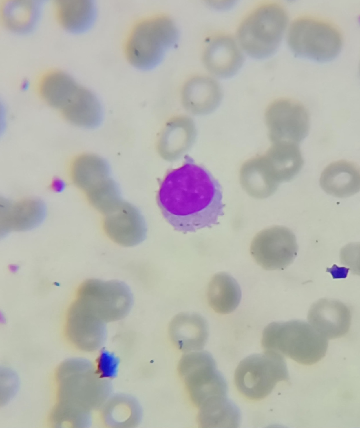
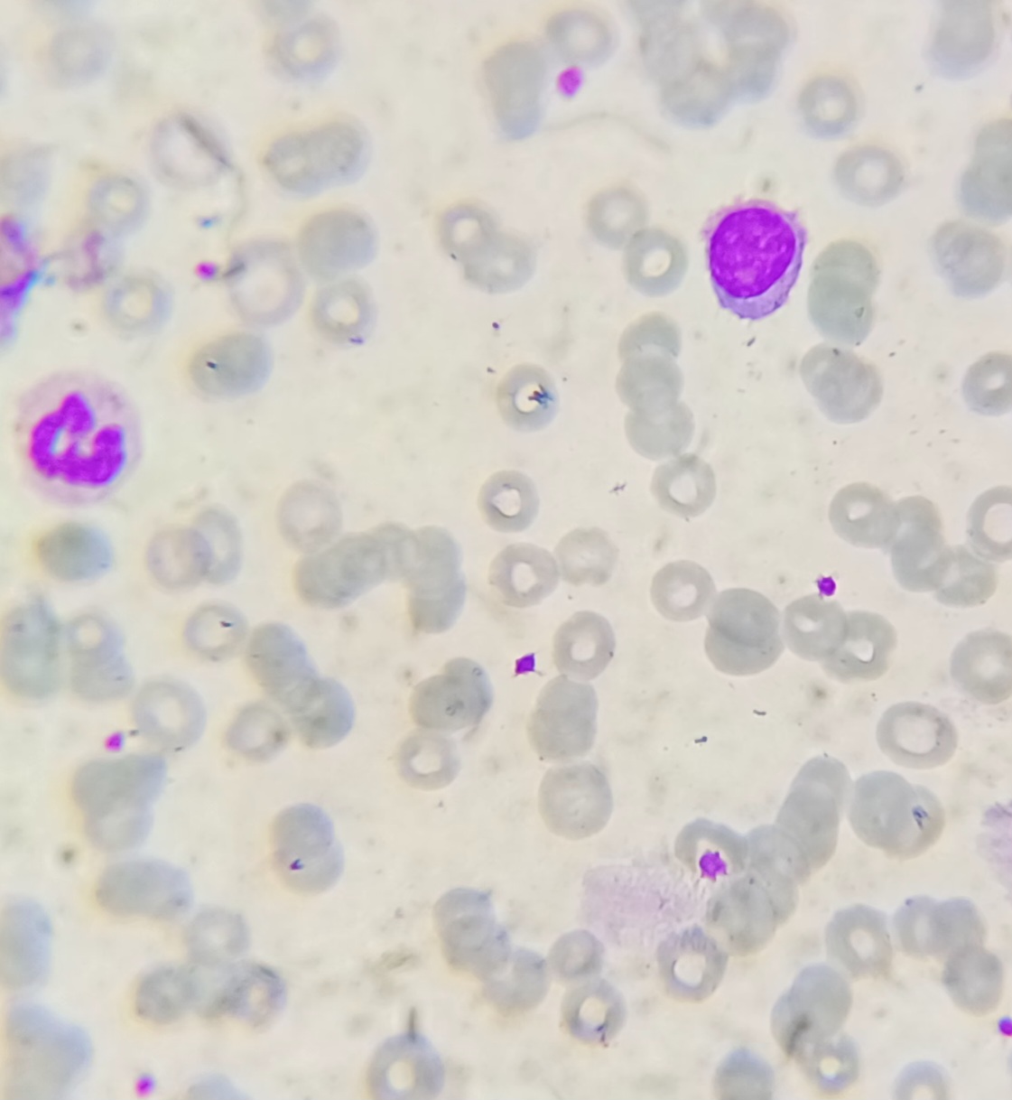
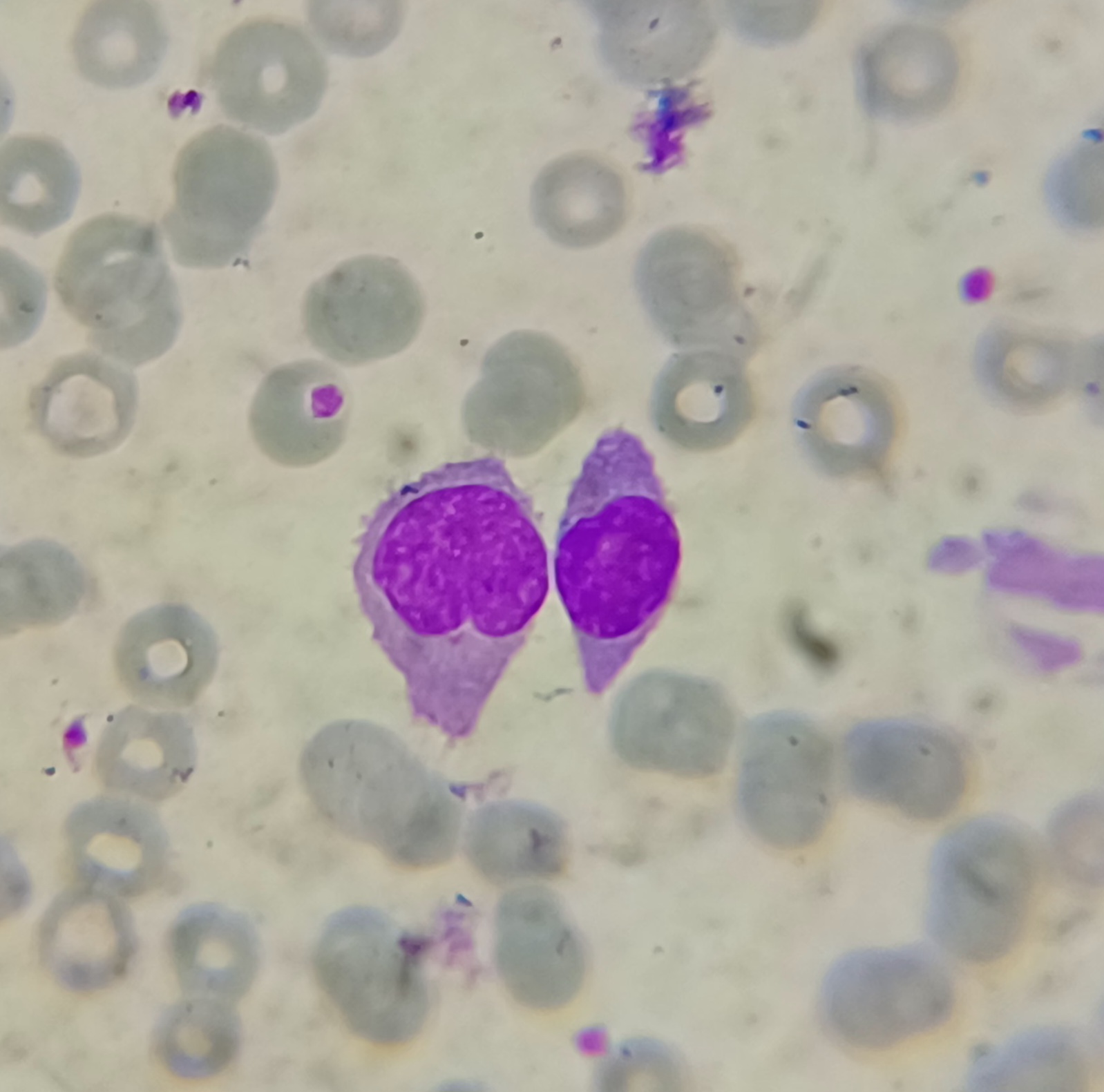
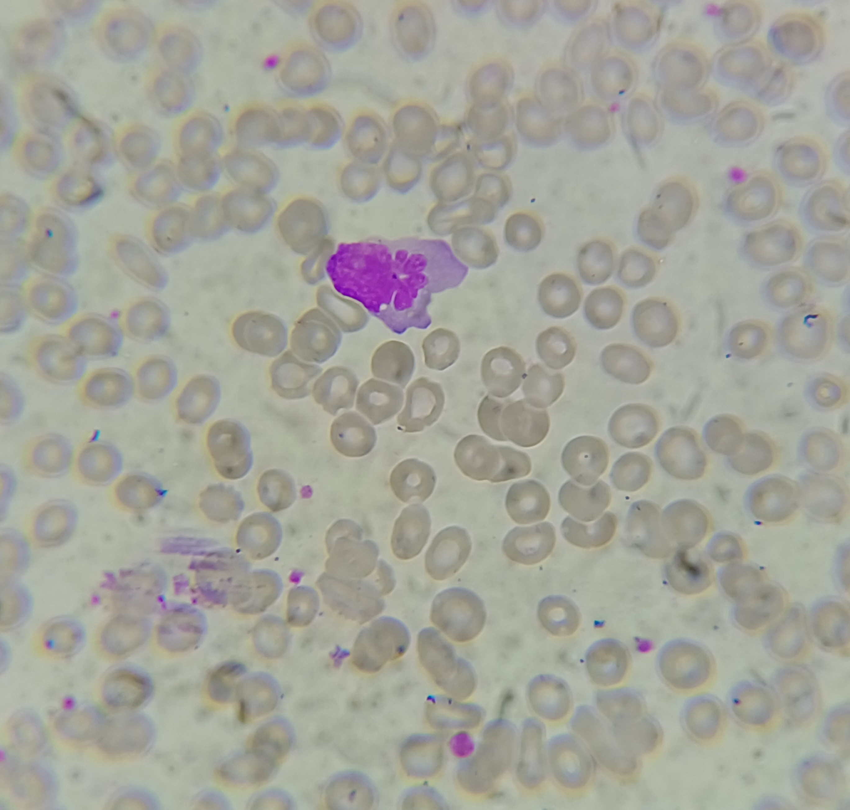
🔬 Peripheral Smear Findings: Look at the images below !!!
🧠 Residents & pathologists—please share your interpretation, differentials, and approach.
📌 Feel free to comment on morphological clues that guide your diagnosis.
20 April 2026: Answer (See IMAGE below)
Differentiating Villous Lymphocytes, Hairy Cells, and Artifact Mimicking Projections


RBC are normocytic with few microcytes.
The lymphocytes look atypical with small to intermediate size and circumferential cytoplasmic projections.
A bit more history required is about splenomegaly status.
Differential diagnosis :-
1) Hairy cell Leukemia
2) Splenic Marginal zone lymphoma
Advice- Flowcytometry evaluation for characterization.
Good approach Dr Prateek
Hairy cell leukemia
Atypical lymphoid cells some with hairy projections
? Hairy cell leukemia
Hairy cell leukemia
T cell leukaemia/ lymphoma spill over
1] RBCs distinctly green in color which means magenta colour is less expressed in these pictures
2] less magenta means the nuclei tend to look more pink and hence look more immature
3] one cell has hairy projections [ likely drying artefact as some RBC s also show artefact ]
other has plenty of cytoplasm and plasmacytoid features one lobate nucleus
and one with monocytoid features
Different cell population [ non uniform ] with dirty blue cytoplasm even when majenta is less STRONGLY FAVORS REACTIVE CHANGE.
Hairy cell leukemia
Good approach Dr Ajit.
Unilineage cytopenia in RBC series. Rouleaux formation in RBCs. Plasmacytoid lymphocytes/ reactive lymphocytes, mocytoid cell with bluish cytoplasm, atypical nucleated cell with plasmacytoid cytoplasm. Also noted is one lymphocyte with hairy projections.
Some cytoplasmic bodies are seen. Pain abdomen could be due to splenomegaly or mass in abdomen . Possibilities include reactive lymphocytes seen in infections, solitary plasmcytoma, Multiple myeloma, plasma cell leukemia and Hairy cell leukemia.
Dr Ashish as this is a peripheral smear we cannot have differentials like solitary plasmacytoma or Multiple Myeloma.Good observation
Hairy call Leukemia
Rouleaux formation with plasmacytoid cells
Activated / Reactive lymphocytes
last pic- Monocyte
Hairy Cell Leukaemia.
Microcytic Hypochromic Anaemia
Hairy cell Leukaemia
Hairy cell leukemia
Hairy cell leukaemia