Dr. Smitirupa Mishra , Consultant Pathologist, Sparsh Hospital , Bhubaneswar, Odisha
MCQ 1:
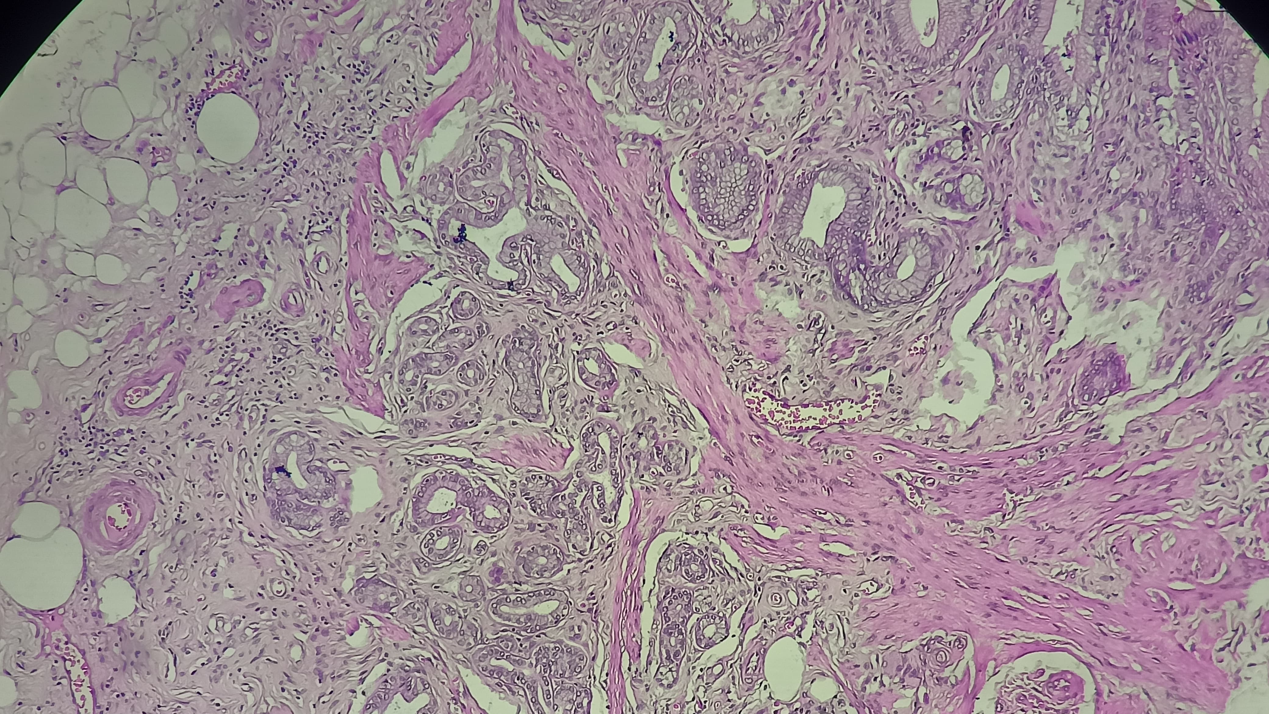
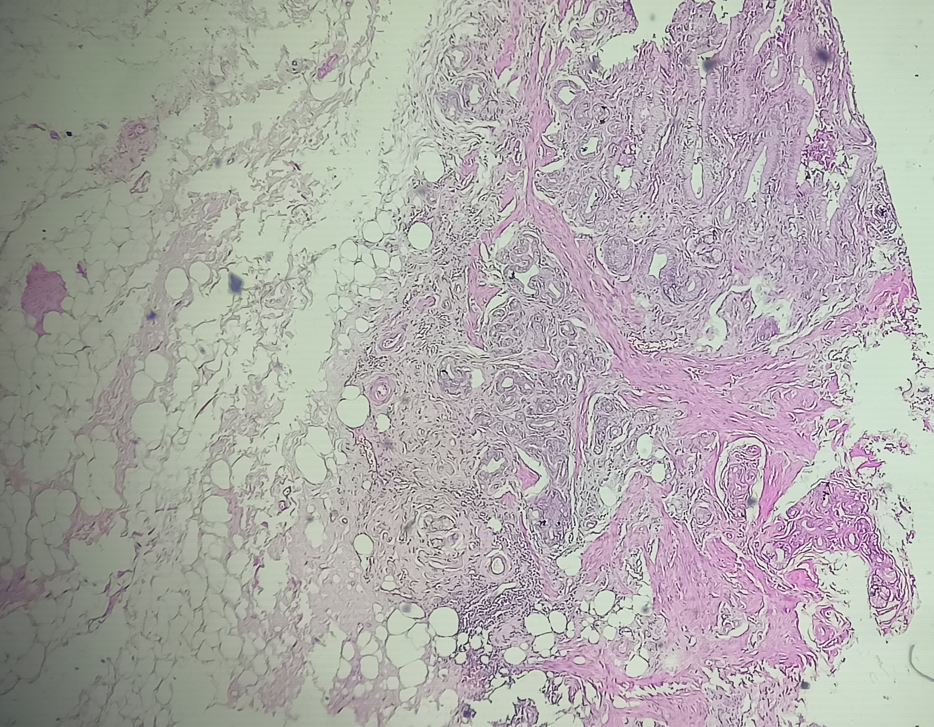
A histopathology image of the gallbladder shows thickened muscular wall with multiple irregular glands extending deep into the muscle layer . What is the most likely diagnosis?
A. Xanthogranulomatous cholecystitis
B. Gallbladder adenocarcinoma
C. Adenomyomatosis of gallbladder
D. Cholesterolosis
E. Acute cholecystitis
MCQ 2:
Which of the following best explains the pathogenesis of the above diagnosed condition?
A. Neoplastic proliferation of glandular epithelium
B. Increased intraluminal pressure causing mucosal herniation into hypertrophied muscle
C. Autoimmune destruction of gallbladder wall
D. Infectious granulomatous inflammation
E. Congenital absence of muscular layer
MCQ 3:
The diagnosis its most commonly associated with:
A. Acute viral hepatitis
B. Chronic cholecystitis
C. Primary sclerosing cholangitis
D. Choledochal cyst
MCQ 4:
What is the clinical significance of this condition?
A. Always progresses to carcinoma
B. Premalignant lesion
C. Benign condition with minimal malignant potential
D. Infectious etiology
E. Requires chemotherapy
ANSWERS (Published 11 May 2026):
Mcq 1- C
Mcq 2- B
Mcq 3- B
Mcq 4- C

1-C
2-B
3-B
4-C
1. (C) Adenomyomatosis of GB
2. (B) Increased intraluminal pressure causing mucosal herniation into hypertrophied muscle.
3.(B).Chronic cholecystitis
4.(C).Benign condition with minimal malignant potential
1-C
2-B
3-B
4-C
MCQ 1. C
MCQ 2. B
MCQ 3. B
MCQ 4. B
MCQ 1: C. Adenomyomatosis of gallbladder
MCQ2: B. Increased intraluminal pressure causing mucosal herniation into hypertrophied muscle
MCQ3: B. Chronic cholecystitis
MCQ4:C. Benign condition with minimal malignant potential
1. C
2.B
3.B
4. C
MCQ 1- C
MCQ 2- B
MCQ 3- B
MCQ 4- C
MCQ
1-C,
2-B,
3-C,
4-C.
C,B,B,C
1. C
2. B
3. B
4. C
1c, 2b, 3b, 4c
1. C
2. B
3. B
4. C
1-c
2-b
3-b
4-c
1.C
2.B
3.B
4.C